Logo
  • contact@literaturepublishers.org
  • Whitley Bay, NE26 2HU, England, United Kingdom
  • Open Access
Article Details
Clinical Image
Volume 4, Issue 4 (April Issue)

A Unique Case of a Large Left-Sided Pericardial Cyst

Kabeer Ali1* and Sateesh Sakhamuri2

1House Officer, Internal Medicine, Medical Associates Hospital, Trinidad and Tobago

2Consultant, Internal Medicine and Pulmonology, Medical Associates Hospital, Trinidad and Tobago

*Corresponding author: Kabeer Ali, House Officer, Internal Medicine, Medical Associates Hospital, Trinidad and Tobago. E-mail: kabeer.ali@hotmail.com

Received: April 23, 2022; Accepted: May 02, 2022; Published: May 18, 2022

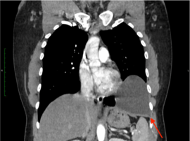
Citation: Ali K, Sakhamuri S, et al. A Unique Case of a Large Left-Sided Pericardial Cyst. Clin Image Case Rep J. 2022; 4(4): 233.

Article Image
Abstract

A 41-year-old male of Afro-Caribbean ancestry presented with a one-year history of dry cough and left lower-sided chest discomfort. He had no known chronic medical conditions. Erect chest X-ray showed an ovoid homogeneous radiodense lesion at the left cardiophrenic angle, and a contrast CT scan of the chest (Figure) confirmed a non-enhancing unilocular cyst abutting pericardium with homogenous fluid density (33 Hounsfield units). An echocardiogram showed a cystic mass and a minimal pericardial effusion. Pericardial cysts are usually right-sided, unilocular, and form due to anomalous nonunion of embryologic pericardial lacunae. They are generally asymptomatic and found incidentally. Mediastinal fat, Morgagni hernia, and bronchogenic cyst are the standard differentials. Surgical excision is indicated if the patient develops chest pain, tamponade, or features of malignancy. An ultrasound-guided percutaneous aspiration was performed as per the patient’s priority, following which he achieved symptomatic relief. The cytology from the aspiration was benign.