
Clinical Images and Case Reports Journal | PubMed
  • contact@literaturepublishers.org
  • Whitley Bay, NE26 2HU, England, UK
  • Submit Manuscript
Article Details

Clinical Image

Volume 3, Issue 8 (August Issue)

Recurrent Pancreatitis Due to an Intraluminal Duodenal Diverticulum

Tomohito Shinoda, Tanahashi Toshiyuki* and Takuya Nakashima

Department of Surgery, Gifu Municipal Hospital, Kashimacho, Gifu, Japan

*Corresponding author: Toshiyuki Tanahashi, Department of Surgery, Gifu Municipal Hospital, Kashimacho, Gifu, Japan. E-mail: c2001049@yahoo.co.jp

Received: August 30, 2021; Accepted: September 13, 2021; Published: September 28, 2021

Citation: Shinoda T, Toshiyuki T, Nakashima T, et al. Recurrent Pancreatitis Due to an Intraluminal Duodenal Diverticulum. Clin Image Case Rep J. 2021; 3(8): 183.
Recurrent Pancreatitis Due to an Intraluminal Duodenal Diverticulum
Abstract

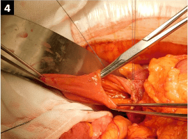
A 35-year-old woman, with a history of multiple admissions for acute pancreatitis since the age of 17 years, visited our hospital with a chief complaint of epigastric pain. She was diagnosed with intussusception of the upper small intestine based on abdominal contrast-enhanced computed tomography findings (Figure 1). Laboratory data were as follows: white blood cell count, 13930/?L; amylase level, 587 U/ L; and C-reactive protein level, 0.24 mg/dL. She was hospitalised for further examination and treatment. Upper gastrointestinal (UGI) endoscopy showed a diverticulum that contained food in the descending duodenum (Figure 2). A UGI contrast study revealed a barium-filled sac within the duodenum (Figure 3); therefore, a diagnosis of an intraluminal duodenal diverticulum (IDD) was made. Mechanical stimulation due to distension caused by food was thought to have caused both the intussusception and the patient’s previous episode of pancreatitis. The diverticulum, which emerged from the duodenal lumen below the papilla of Vater, was surgically removed to prevent relapse of the conditions (Figure 4). The resected diverticulum was covered with a mucous membrane, and the inherent muscular layer was lost in the diverticulum (Figure 5). The patient had no symptoms postoperatively.

IDD is a rare congenital anomaly [1]. Its nonspecific symptoms make diagnosis difficult and necessitate appropriate imaging [2]. The ‘windsock sign’ is typical in a UGI contrast study of a patient with IDD [2,3]. IDD should be suspected when patients present with recurrent epigastric pain and pancreatitis of unknown aetiology.

hacklink satın al coinbar coinbar giriş grandpashabet grandpashabet giriş grandpashabet güncel giriş grandpashabet grandpashabet giriş grandpashabet giriş güncel alobet alobet giriş betcio betcio giriş betcio betcio giriş holiganbet giriş holiganbet jojobet jojobet giriş meritking meritking giriş madridbet madridbet giriş meritking giriş meritking pashagaming pashagaming giriş jojobet güncel giriş holiganbet holiganbet giriş holiganbet holiganbet giriş holiganbet güncel giriş best online casinos in new zealand casibom casibom giriş casibom güncel giriş grandpashabet grandpashabet giriş grandpashabet güncel giriş casibom casibom giriş casibom güncel giriş betcio betcio giriş alobet alobet giriş atlasbet atlasbet giriş pashagaming pashagaming giriş pashagaming pashagaming giriş madridbet madridbet giriş kingroyal kingroyal giriş holiganbet holiganbet giriş grandpashabet grandpashabet giriş grandpashabet güncel giriş holiganbet holiganbet giriş holiganbet güncel giriş casibom casibom giriş casibom güncel giriş meritking meritking giriş meritking güncel giriş holiganbet betbey esbet holiganbet jojobet holiganbet jojobet giriş holiganbet 1win cashwin betpuan sonbahis grandpashabet casinoroyal grandpashabet jasminbet jasminbet giriş