Clinical Images and Case Reports Journal | PubMed
  • contact@literaturepublishers.org
  • Whitley Bay, NE26 2HU, England, UK
  • Submit Manuscript
Article Details

Clinical Image

Volume 3, Issue 6 (June Issue)

Vascular Sheathing from Ischemic Central Retinal Vein Occlusion

Michael L Marin1* and Adam Korayem2

1Surgical Department, Icahn School of Medicine at Mount Sinai, USA
2Vascular Surgery Department, Icahn School of Medicine at Mount Sinai, USA

*Corresponding author: Michael L Marin, Surgical Department, Icahn School of Medicine at Mount Sinai, USA, E-mail: martin.michael@gmail.com

Received: May 28, 2021; Accepted: June 06, 2021; Published: June 19, 2021

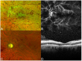
Citation: Marin ML, Korayem A, et al. Vascular Sheathing from Ischemic Central Retinal Vein Occlusion. Clin Image Case Rep J. 2021; 3(6): 168.

Vascular Sheathing from Ischemic Central Retinal Vein Occlusion
Abstract

A 54-year-old man with no prior medical care presented with acute vision loss in the left eye. Visual acuity was hand motion and blood pressure were 220/136. Examination demonstrated severe disc and macular edema, retinal whitening, venous sheathing, and intraretinal hemorrhages (Panel A). Fluorescein angiography revealed venous leakage and non-perfusion (Panel B). The patient underwent empiric intravitreal foscarnet. After infectious/inflammatory etiologies were ruled out, he received 3 monthly intravitreal bevacizumab injections for presumed central retinal vein occlusion. At 3 months, there was complete resolution of sheathing (Panel C) but with persistent macular ischemia (Panel D) resulting in counting fingers vision.