Clinical Images and Case Reports Journal | PubMed
  • contact@literaturepublishers.org
  • Whitley Bay, NE26 2HU, England, UK
  • Submit Manuscript
Article Details

Clinical Image

Volume 4, Issue 5 (May Issue)

A Bullet in the Thoracic Spine and May-Thurner Syndrome

Elior Moreh1,2*, Sheer Shabat1, Nurith Hiller2,3 and Efrat Suraqui1

1Physical Medicine and Rehabilitation Department, Hadassah Hebrew University Medical Center, Jerusalem, Israel

2Faculty of Medicine, Hebrew University, Jerusalem, Israel

3Radiology, Hadassah Hebrew University Medical Center, Jerusalem, Israel

*Corresponding author: Elior Moreh, Physical Medicine and Rehabilitation Department, Hadassah Hebrew University Medical Center, POB 24035, Jerusalem 92140, Israel. E-mail: elior@hadassah.org.il

Received: May 12, 2022; Accepted: May 20, 2022; Published: June 03, 2022

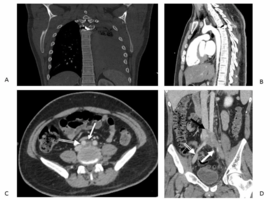
Citation: Moreh E, Shabat S, Hiller N, Suraqui E, et al. A Bullet in the Thoracic Spine and May-Thurner Syndrome. Clin Image Case Rep J. 2022; 4(5): 234.

A Bullet in the Thoracic Spine and May-Thurner Syndrome
Abstract

A 24-year-old man was treated in the rehabilitation department following complete paraplegia due to a gunshot injury from a bullet that entered through the left scapula via the left lung and remained stuck in the body of his T4 vertebra (Panel A – Computerized Tomography showing the bullet bulging from the body of T4 vertebra into the spinal cord, Panel B shows massive left hemothorax next to the bullet). He had flaccid muscle tone in his legs and was wheelchair-ridden.

Despite preventive treatment with enoxaparin 60 mg a day, on day 45 after the injury he developed acute swelling of his left leg, including the shin and the thigh. The leg was warm and firm but not tender since the patient had no sensation below the T5 level. A Doppler Ultrasound of the lower extremities revealed he had a deep venous thrombosis (DVT) of the left common iliac vein. Computerized Tomography Venography revealed an anatomical variant predisposing him for left DVT: an overlying right common iliac artery (parallel white arrow, panels C and D compressing the left common iliac vein (solid white arrow, panels C and D). The clot occluding the left common iliac vein extends into the Inferior Vena Cava (black arrow, Panel D). This is the classical anatomical variant seen in May-Thurner syndrome – a rare condition in which compression of the common venous outflow tract of the left lower extremity leads to leg swelling, pain, and eventually blood clots. Enoxaparin dose was increased to 60 mg twice a day and the swelling of the left leg resolved almost completely. The patient achieved full independence in transfers and wheelchair mobility and was instructed to stop enoxaparin treatment 6 months after the occurrence of the DVT.