Clinical Images and Case Reports Journal | PubMed
  • contact@literaturepublishers.org
  • Whitley Bay, NE26 2HU, England, UK
  • Submit Manuscript
Article Details

Clinical Image

Volume 3, Issue 3 (March Issue)

Acute Blindness After Lower Limb Injury

L. Jay-Caillierez1*, B. Allaouchiche2 and F. Cotton3

1Lucille Jay-Caillierez, Hospices Civils de Lyon, Service d’Anesthésie Réanimation, Groupement Hospitalier Sud, F-69495, Pierre Bénite, France
2Bernard Allaouchiche, Hospices Civils de Lyon, Service d’Anesthésie Réanimation, Groupement Hospitalier Sud, F-69495, Pierre Bénite, France
3François Cotton, Hospices Civils de Lyon, Service de Radiologie, Groupement Hospitalier Sud, F-69495, Pierre Bénite, France

*Corresponding author: Lucille Jay-Caillierez, Hospices Civils de Lyon, Service d’Anesthésie Réanimation, Groupement Hospitalier Sud, F-69495, Pierre Bénite, France, Tel: +33478861476; E-mail: lucille.jay@chu-lyon.fr

Received: March 30, 2021; Accepted: April 07, 2021; Published: April 28, 2021

Citation: L. Jay-Caillierez, B. Allaouchiche, F. Cotton, et al. Acute Blindness After Lower Limb Injury. Clin Image Case Rep J. 2021; 3(3): 155.

Acute Blindness After Lower Limb Injury
Abstract

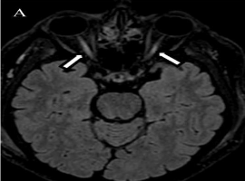
Key Points: A patient with complex comminutive fracture of leg presents bilateral blindness. The brain MRI showed bilateral optic nerves hypersignal on FLAIR and diffusion sequences, suggesting a diagnosis of fat embolism.

Clinical Image
A 57-years old man was treated in a trauma center after a motorcycle accident. He had a complex comminuted fracture of the left leg with acute limb ischemia. A popliteal-tibia bypass was performed in emergency and an external fixator was implanted. The intraoperative period was marked by a major instability due to hemorrhagic shock and reperfusion syndrome, requiring massive transfusion and high doses of vasopressor. The next day, the patient reported a bilateral blindness upon waking up; the rest of the patient's neurological examination was normal without other focal deficit. The brain computed tomographic scan was strictly normal. The brain MRI performed four days after the trauma showed bilateral optic nerves hypersignal on both FLAIR and diffusion sequences (A, B). Two diagnostics were discussed: a fat embolism and an ischemic posterior neuropathy, which has been reported following hypovolemic shock. The most likely diagnosis remains fat embolism in this context, considering acute lesions in basal ganglia sparing the junctional territories suggesting an embolism mechanism [1-3]. Of note the blood lipid changes seen at day 5 following the trauma including, with a decrease in cholesterol level (1,29 g/l) and increase in triglycerides values (2,18 g/l), which have been described in fat embolism [4]. Unfortunately, no bronchoalveolar lavage has been performed. The funduscopic exam performed 2 days after the accident was normal, without emboli or hemorrhage and a clear and colored papilla but no embolus. To date (1 year later), the patient has not recovered visual acuity.