Clinical Images and Case Reports Journal | PubMed
  • contact@literaturepublishers.org
  • Whitley Bay, NE26 2HU, England, UK
  • Submit Manuscript
Article Details

Clinical Image

Volume 3, Issue 2 (February Issue)

Atypical Location of Tophaceous Gout

Ana Oliveira Monteiro1*, Helena Barroca2 and M Teresa Cardoso3

1Department of Internal Medicine, Centro Hospitalar Universitario de Sao Joao, Porto, Portugal
2Department of Pathology, Centro Hospitalar Universitario de Sao Joao, Porto, Portugal
3Department of Internal Medicine, Centro Hospitalar Universitario de Sao Joao, Porto, Portugal

*Corresponding author: Ana Oliveira Monteiro, Department of Internal Medicine, Centro Hospitalar Universitario de Sao Joao, Porto, Portugal, Tel: +351225512200; E-mail: ana.galraoom@gmail.com

Received: January 13, 2021; Accepted: January 22, 2021; Published: February 24, 2021

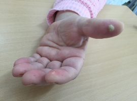
Citation: Oliveira Monteiro A, Barroca H, Cardoso MT, et al. Atypical Location of Tophaceous Gout. Clin Image Case Rep J. 2021; 3(2): 142.

Atypical Location of Tophaceous Gout
Abstract

A 73-years-old female with a past medical history of obesity, dyslipidemia, type 2 diabetes with nephropathy and chronic kidney disease stage G3A1 (KDIGO classification), hyperuricemia and dyslipidemia was admitted to the hospital with severe dehydration, worsening kidney function and lactic acidosis related to acute watery diarrhea and maintenance of therapy with metformin, furosemide and non-steroidal anti-inflammatory drugs, needing transient hemodialysis. She presented polyarthritis of the small joints of the left hand, and several hard white nodules surrounded by an inflamed halo at the fingertips (Figure 1A and B).

The main lesion was drained and histology revealed abundant urate crystals (Figure 2), confirming the clinical diagnosis of tophaceous gout. Treatment with colchicine was started, with clinical improvement.
Gout is a clinical syndrome, characterized by monosodium urate crystal deposition [1]. Hyperuricemia (saturation of serum for urate) is the precondition for the deposit of the urate crystals, but most patients never experience a clinical event. The natural history of gout occurs in four stages: asymptomatic hyperuricemia; acute gout flares; intercritical gout; and chronic gouty arthritis with tophaceous gout. Tophi are the most characteristic lesions of gout, characterized by deposits of monosodium urate crystals, accompanied by inflammation in the surrounding tissue [1]. Lesions are usually palpable, firm, white, or yellow and typically present in the ears, or articular structures, tendons, or bursas. The usual presentation evolves a single joint, the most common is the first metatarsophalangeal or knee. In some cases, as in our case report, tophi can present in atypical locations. This is particularly rare and usually occurs in women, elderly, and transplants recipients [2]. Tophi can form in any area containing connective tissue, and atypical places of presentation are related in the literature to chronic kidney disease and treatment with diuretics or anti-inflammatory drugs, as in our case report [3].