Clinical Images and Case Reports Journal | PubMed
  • contact@literaturepublishers.org
  • Whitley Bay, NE26 2HU, England, UK
  • Submit Manuscript
Article Details

Clinical Image

Volume 4, Issue 7 (July Issue)

Diag-nose-ing Septic Emboli

Tony Zhang1* and Peter Alan Barber2

1Neurology Registrar, Department of Neurology, Auckland City Hospital, 2 Park Road, Grafton, Auckland 1023, New Zealand

2Professor of Neurology, Department of Neurology, Auckland City Hospital, 2 Park Road, Grafton, Auckland 1023, New Zealand

*Corresponding author: Tony Zhang, Neurology Registrar, Department of Neurology, Auckland City Hospital, 2 Park Road, Grafton, Auckland 1023, New Zealand. E-mail: tonyz@adhb.govt.nz

Received: June 29, 2022; Accepted: July 07, 2022; Published: July 20, 2022

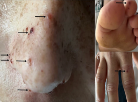
Citation: Zhang T, Barber PA. Diag(nose)ing Septic Emboli. Clin Image Case Rep J. 2022; 4(7): 247.

Diag-nose-ing Septic Emboli
Abstract

A 27-year old previously well female presented after waking with right lower facial droop and dysarthria. Over the previous month, she complained of fatigue, anorexia and night sweats. The patient noticed new “spots” on her nose that she initially thought were freckles (Figure A). Examination also revealed concurrent Janeway’s lesions in the toes (Figure B) and petechial lesions in the fingers (Figure C) consistent with peripheral stigmata of infective endocarditis. A loud pan-systolic murmur was audible at the mitral region. Oral examination revealed poor dentition.

Blood cultures were positive for streptococcus mitis. Transoesophageal echocardiogram revealed a 3.4 x 2.8cm vegetation attached to the posterior mitral valve leaflet. Non-contrast CT head confirmed an embolic infarct in the left frontal lobe involving the precentral gyrus.

Intravenous ceftriaxone was commenced, and she underwent successful mitral valve repair. Follow up six weeks later revealed complete resolution of the embolic skin lesions on her nose. A diagnosis of septic embolization to the nose was made, which has not previously been described in the literature. This serves as a reminder for clinicians to look beyond the peripheries for stigmata of infective endocarditis.

Keywords: Septic emboli; Stroke; Infective endocarditis; Embolic skin lesions