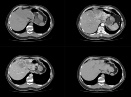
A man in his early sixties underwent abdominal ultrasound (Figure 1) for unrelated reasons, which incidentally demonstrated marked structural heterogeneity of the left hepatic lobe. Contrast-enhanced computed tomography (Figure 2,3) revealed replacement of the entire left lobe by multiple confluent lesions showing peripheral globular enhancement with progressive centripetal filling on delayed phases. This pattern suggested a diffuse vascular process rather than a single heterogeneous mass. Several additional nodules were identified in the right hepatic lobe, displaying the classic enhancement pattern of sporadic hepatic hemangiomas. A 5 cm exophytic lesion adjacent to segment II, possibly pedunculated, was also noted.
Magnetic resonance imaging (MRI) (Figure 4,5) confirmed multiple confluents, T2-hyperintense nodular lesions, with centripetal filling, predominantly involving the left lobe, extending approximately 15 × 10 cm in the axial plane. Additional smaller nodules in the right lobe showed identical enhancement characteristics. The overall constellation of findings was highly suggestive of diffuse hepatic hemangiomatosis, a rare benign vascular condition characterized by the proliferation of numerous hemangiomatous lesions that may coalesce and involve large regions of hepatic parenchyma.
Importantly, hepatic hemangiomatosis differs from sporadic hemangiomas in both distribution and behavior. Sporadic hemangiomas are typically solitary or few, well-circumscribed, and confined to limited regions of the liver. In contrast, hemangiomatosis is characterized by a large number of lesions—often confluent—sometimes replacing entire lobes, as in this case. Unlike hemangiomas, which rarely distort hepatic architecture, hemangiomatosis can lead to diffuse parenchymal alteration while still preserving lobar morphology. The distinction is clinically relevant, as extensive hemangiomatosis may raise concern for infiltrative disease on initial imaging.
Key Message: Recognition of the characteristic centripetal enhancement pattern across multiple lesions is essential to differentiate diffuse hepatic hemangiomatosis from sporadic hemangiomas and from infiltrative malignant processes. MRI remains the optimal modality for confident noninvasive diagnosis and to prevent unnecessary biopsy or intervention.
")
")