Clinical Images and Case Reports Journal (CICRJ)
  • contact@literaturepublishers.org
  • Whitley Bay, NE26 2HU, England, UK
  • Submit Manuscript
Article Details

Case Report

Volume 8, Issue 3

Dynamic Fluoroscopic Recognition of Chronic Subclavian Vein Occlusion During CIED Implantation: Mechanobiological Remodeling and Functional Venous Patency

Abdullah Alabdulgader*

Congenital Cardiologist, Electrophysiologist, Saudi Arabia

*Corresponding author: Abdullah Alabdulgader, MD, DCH (Dublin), DCH (Edinburgh), MRCP (UK), ABP (CAMH), FRCP (Edinburgh), Pacing and Electrophysiology (University of Alberta-Canada). Senior Scientist, Congenital Cardiologist, interventional electrophysiologist and cardiac rhythm devices implanter, Psychophysiologist, Philosopher, World Gold Medal Awardee (Wosco-2012). Scientific Advisory Board Member (Heart Math Institute-USA).
E-mail: Alabdulgader.ep@gmail.com

Received: May 14, 2026; Accepted: June 01, 2026; Published: June 15, 2026

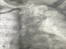
Citation: Alabdulgader A. Dynamic Fluoroscopic Recognition of Chronic Subclavian Vein Occlusion During CIED Implantation: Mechanobiological Remodeling and Functional Venous Patency. Clin Image Case Rep J. 2026; 8(3): 595.

Dynamic Fluoroscopic Recognition of Chronic Subclavian Vein Occlusion During CIED Implantation: Mechanobiological Remodeling and Functional Venous Patency
Abstract

Chronic thoracic venous obstruction is an increasingly encountered but frequently underrecognized challenge during cardiac implantable electronic device (CIED) implantation. Because progressive collateral venous development may preserve effective upper extremity drainage, severe venous obstruction can remain entirely asymptomatic and become apparent only during attempted transvenous instrumentation. We report a case of incidentally discovered chronic total left subclavian vein occlusion identified during permanent pacemaker implantation in a patient without clinical manifestations of venous obstruction. Initial left-sided venous access demonstrated successful aspiration of venous blood; however, fluoroscopic guidewire advancement revealed an abnormal superior and lateral trajectory inconsistent with normal central venous anatomy. Subsequent contrast venography confirmed chronic total left subclavian vein occlusion with extensive collateral venous circulation and delayed contralateral venous opacification. Following abandonment of the left-sided approach, contralateral right subclavian venography demonstrated an angiographically narrowed and medially tapered venous segment that initially raised concern for additional obstruction. Nevertheless, careful dynamic fluoroscopic assessment confirmed preserved functional luminal continuity, permitting successful right ventricular lead implantation without complications. This case highlights several important procedural and mechanobiological principles relevant to contemporary electrophysiology practice, including the remarkable adaptive capacity of thoracic collateral venous remodeling, the limited reliability of venous blood aspiration alone in confirming central venous patency, and the critical importance of abnormal guidewire trajectory as an early fluoroscopic marker of occult venous obstruction. Furthermore, the case emphasizes the distinction between static angiographic appearance and true functional venous traversability. Early recognition, procedural adaptability, and meticulous fluoroscopic assessment remain essential for safe and successful management of complex venous anatomy during CIED implantation.