Clinical Images and Case Reports Journal | PubMed
  • contact@literaturepublishers.org
  • Whitley Bay, NE26 2HU, England, UK
  • Submit Manuscript
Article Details

Clinical Image

Volume 7, Issue 6

Falciform Ligament Necrosis: A Rare Clinical Entity

Pretty Sara Idiculla*, Vinay Shet and Jobin Philipose

Mountain View Regional Medical Center, 4311 E Lohman Ave, Las Cruces, New Mexico

*Corresponding author: Pretty Sara Idiculla, MD, Mountain View Regional Medical Center, Las Cruces, New Mexico. E-mail: sarahidiculla.psi@gmail.com

Received: August 25, 2025; Accepted: September 08, 2025; Published: September 15, 2025

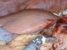
Citation: Idiculla PS, Shet V, Philipose J. Falciform Ligament Necrosis: A Rare Clinical Entity. Clin Image Case Rep J. 2025; 7(6): 575.

Falciform Ligament Necrosis: A Rare Clinical Entity
Abstract

A 39-year-old male with past medical history of fatty liver disease, Helicobacter pylori associated peptic ulcer disease, and migraines, presented for sharp, progressively worsening RUQ pain. History of similar episodes in the past that usually lasts less than 1 hour. The pain radiated to his right shoulder and tenderness was appreciated in the RUQ. USG showed gallbladder wall thickening with a positive sonographic Murphy sign, and no gall stones. CT scan was unremarkable and HIDA scan showed gallbladder EF of 5% (biliary dyskinesia). Treatment started with intravenous cefepime, no improvement and as per General Surgery recommendations, he was taken for robotic cholecystectomy. Intraoperatively, omental adhesion to the falciform ligament with hemorrhagic changes and evidence of inflammation indicating necrosis was noted. Gall bladder appeared normal and was removed. Pathology revealed hemorrhagic fat necrosis of Falciform ligament and minor evidence of chronic cholecystitis with cholesterolosis. There was immediate post-surgical clinical improvement, no noted complications, and patient was discharged on oral antibiotics to complete a 7-day course.

Falciform ligament necrosis is a rare diagnosis and can mimic the presentation of cholecystitis, pancreatitis, or perforated peptic ulcer. Our case is unique as the patient showed no imaging findings indicative of falciform ligament necrosis and failed conservative management. Even though acute cholecystitis is a commonly encountered pathology, this report indicates the importance of broadening differentials in patients who present with atypical symptoms or normal imaging findings but show no clinical improvement with conservative management. In these cases, it would be important to involve multidisciplinary teams that involve possibly surgical intervention for definitive diagnosis and treatment.