An 84-year-old female, genetically diagnosed with late-onset Pompe disease (LOPD), gradually presented with swelling on the right side of her neck during the inspiratory phase on non-invasive positive pressure ventilation (NPPV).
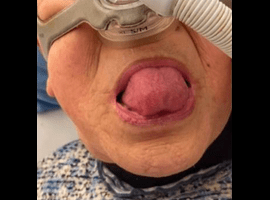
She required NPPV during day and night in her 70s with high end-expiratory positive airway pressure of 15 hPa, because of upper airway obstruction due to macroglossia (Figure 1A).
At her age of 82 years old, she noticed swelling on the right side of her neck during inspiratory phase on NPPV. Over two years, neck swelling was gradually increasing in size.
On physical examination, swelling on the right side of her neck was detected during inspiratory phase via nose, and rapidly returned at expiratory phase via mouth (Video 1), but slightly decreased in size via nose (Video 2).
Clinical Video 1: Inspiratory via nose, and expiatory via mouth. Swelling on the right side of her neck was detected during inspiratory phase, and disappeared at expiratory phase via mouth.
Clinical Video 2: Both of inspiratory and expiatory via nose. Similarly, swelling appeared on her neck during inspiratory phase, but slightly decreased in size and remained swollen even at expiratory phase via nose.
Plain posteroanterior radiographs at inspiratory and expiratory phase demonstrated an air-filled mass in the right, anterior, paramedian region (Figure 1B and 1C). Neck CT revealed severe atrophy of neck muscles, compared to control patient (Figure 2A and 2C). Even in expiratory phase, distended pyriform sinus with air-filled outpouching was observed (Figure 2A and 2D) and further extended across the thyrohyoid membrane inspiratory phase was detected (Figure 2B and 2E), compared to control (Figure 2C). A diagnosis of laryngocele, which is the dilation of the laryngeal saccule between false and true vocal cords, was confirmed.
The phenomenon was similar to how a frog expands its throat. Male frogs have a “vocal sac,” a sound-resonating throat pouch. When they exhale with the mouth and nostrils closed, the vocal sac expands as its fills with air [1]. In the present case, the combination of dilated piriform fossae behaving like a “vocal sac” in frogs, increased elasticity of the throat wall with thinning tissue following severe muscle atrophy, and continuously high expiratory pressure due to upper airway obstruction due to macroglossia, may have contributed to this complication. We named this finding the “Frog sign,” and suggest including it among the list of complications that may occur in patients with LOPD receiving NPPV.
Keywords: Frog sign; Late-onset Pompe disease; Throat expansion; Non-invasive positive pressure ventilation
")
")