Clinical Images and Case Reports Journal | PubMed
  • contact@literaturepublishers.org
  • Whitley Bay, NE26 2HU, England, UK
  • Submit Manuscript
Article Details

Clinical Image

Volume 2, Issue 2 (May Issue)

Giant Thoracoabdominal Aortic Aneurysm after EVAR

Calin Pop1* and Sabin Nemes Bota2

1Emergency Clinical County Hospital Baia Mare, “Vasile Goldis” University, Faculty of Medicine Arad, Romania

2Emergency Clinical County Hospital Baia Mare, Romania 
 
*Corresponding author: Calin Pop, Emergency Clinical County Hospital of Baia Mare, George Cosbuc street nr 31, Baia Mare 430110, Romania, Tel: 0040744515794; E-mail: medicbm@yahoo.com 

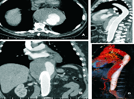
Citation: Calin Pop, Sabin Nemes Bota, et al. Giant Thoracoabdominal Aortic Aneurysm after EVAR. Clin Image Case Rep J. 2020; 2(2): 113.

Giant Thoracoabdominal Aortic Aneurysm after EVAR
Abstract

Endoleak is the most frequent complication and occurs in 10 to 44% of all patients after Endovascular Repair (EVAR) of abdominal aortic aneurysms. We describe the case of an asymptomatic 69-year-old male patient, lost of follow-up, which presents 3 years after EVAR, with a giant thoracoabdominal aortic aneurysm, due to a type IIa endoleak. This asymptomatic patient has an endoleak partially seal by thrombosis but also an important enlargement that may lead to aneurysm rupture. We propose first a percutaneous transarterial embolization with glue (60% success in our series) but despite the knowledge that aneurysm rupture may occur our patient refuse any intervention. Endoleaks treatment is a real clinical challenge and the therapeutic outcomes are still controversial. 
 
A 69-year-old male, asymptomatic, lost of follow-up, presents 3 years after endovascular repair (EVAR) for a 50 mm diameter and 70 mm long thoracoabdominal aortic aneurysm type III. At, that time he received 2 successive MFM (Multilayer Flow Modulator) stents grafts of 40/100 mm and 45/100 mm. Images from CT angiography demonstrate contrast material outside the stent lumen indicating persistent endoleak and partial thrombosis of a huge aneurysm sac 92 mm diameter and 142 mm long (Figure A and B). Reconstructed CT show perigraft blood flow caused by an endoleak type II a (most probably Inferior Mesenteric Arteries-IMA) seen as a blush anterior to the aortic stent graft (Figure C and D).   The incidence of type II endoleaks after EVAR has been reported as between 10% and 44% [1]. A type II endoleak is attributed to retrograde flow from the IMA (IIa), lumbar arteries (IIb), or other collateral vessels. Current guidelines recommend intervention in type II endoleak when expansion of sac diameter ? 10 mm/year, detected during follow up using the same imaging modality and measurement method [2]. Unfortunately, optimal thresholds are not clear. Endovascular techniques are the preferred modality but surgical repair remains the definitive option in cases where less invasive methods have failed or are precluded [3]. Our asymptomatic patient, 3 years after EVAR have a persistent endoleak partially seal by thrombosis, but also an important enlargement (progression more than 10 mm/year) that may lead in our opinion to aneurysm rupture. We propose first a percutaneous transarterial embolization with glue (60% success in our series) by transbrachial or transfemoral access. Despite the knowledge that aneurysm rupture may occur our patient refuse any intervention. Endoleaks treatment is a real clinical challenge and until solutions are found, EVAR will remain an imperfect long-term treatment and continued follow-up will be mandatory.