Clinical Images and Case Reports Journal | PubMed
  • contact@literaturepublishers.org
  • Whitley Bay, NE26 2HU, England, UK
  • Submit Manuscript
Article Details

Clinical Image

Volume 4, Issue 2 (February Issue)

Incidental Finding of Muscle Lymphoma in an Adult with Common Variable Immune Deficiency

Maimaris J1,2*, Wagner T3, Bowcock S4 and Elfeky R1,2

1Department of Immunology, Royal Free London NHS Trust, London, United Kingdom

2Institute of Immunity and Transplantation, University College London (UCL), London, United Kingdom

3Department of Nuclear Medicine, The Royal Free London NHS Trust, London, United Kingdom

4King’s College Hospital NHS Foundation Trust, London, United Kingdom

*Corresponding author: Jesmeen Maimaris, Department of Immunology, Royal Free London NHS Trust; Institute of Immunity and Transplantation, University College London (UCL), London, United Kingdom. E-mail: j.maimaris@ucl.ac.uk

Received: February 24, 2022; Accepted: March 07, 2022; Published: March 25, 2022

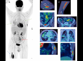
Citation: Maimaris J, Wagner T, Bowcock S, Elfeky R, et al. Incidental Finding of Muscle Lymphoma in an Adult with Common Variable Immune Deficiency. Clin Image Case Rep J. 2022; 4(2): 218.

Incidental Finding of Muscle Lymphoma in an Adult with Common Variable Immune Deficiency
Abstract

A 56-year-old man with common variable immunodeficiency (CVID), granulomatous lymphocytic interstitial lung disease (GLILD) and splenomegaly was reviewed after previous nephrolithotomy stent insertion. CT abdomen found an incidental mesenteric mass which prompted an investigative fluorodeoxyglucose (FDG)- PET-CT scan. MIP image (A) shows two linear areas of intense uptake in muscles of the proximal right thigh (blue arrow), moderate uptake in the mesenteric mass (red arrow) and low-grade uptake in lung changes (green arrows). Fused images of the right thigh (axial B and sagittal C) show intense uptake in muscles of the proximal right thigh. Fused images of the abdomen (coronal D and axial E) show moderate uptake in a nodal left mesenteric mass. Fused images of the lungs (axial F and coronal G) show low-grade uptake in pulmonary fibrosis related to granulomatous lymphocytic interstitial lung disease. Fused images of the left chest wall (axial H and coronal I) show intense uptake in muscles of the left chest wall.

Biopsy confirmed low grade nodal marginal zone lymphoma. Given his asymptomatic presentation and low volume disease, active monitoring is the recommended treatment course. However, given the patient’s concurrent lung disease and multiple neoplastic lesions, rituximab monotherapy is indicated. CVID is the most frequently encountered symptomatic primary immunodeficiency in adults; characterised by hypogammaglobulinemia and recurrent bacterial infections. Patients with CVID are prone to developing autoimmune complications, granulomatous disease and have an increased chance of developing malignancy, particularly lymphoma [1].