Clinical Images and Case Reports Journal | PubMed
  • contact@literaturepublishers.org
  • Whitley Bay, NE26 2HU, England, UK
  • Submit Manuscript
Article Details

Clinical Image

Volume 4, Issue 3 (March Issue)

Migration of a Jugular Hemodialysis Catheter Revealed by Edema of the Left Upper Limb

Hafedh Daly1*, Abdelbaki Bouallegue2 and Amira Horchani3

1Cardiovascular Surgery Department, Regional Hospital of Gafsa, Tunisia 2Nephrology and Hemodialysis Department, Regional Hospital of Gafsa, Tunisia 3Faculty of Pharmacy, Monastir, Tunisia

*Corresponding author: Hafedh Daly, Cardiovascular Surgery Department, Regional Hospital of Gafsa, Tunisia. E-mail: daly.hafedh@yahoo.fr

Received: March 20, 2022; Accepted: March 28, 2022; Published: April 15, 2022

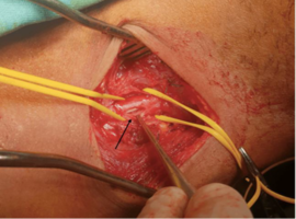
Citation: Daly H, Bouallegue A, Horchani A, et al. Migration of a Jugular Hemodialysis Catheter Revealed by Edema of the Left Upper Limb. Clin Image Case Rep J. 2022; 4(3): 224.

Migration of a Jugular Hemodialysis Catheter Revealed by Edema of the Left Upper Limb
Abstract

A 42-year-old patient, on chronic dialysis for 3 years by a left humero-basilic arteriovenous fistula, consulted for a significant edema of the left upper limb happening for a month (Figure A). The examination found a functional arteriovenous fistula with the presence of multiple small left cervical scars synonymous with iterative placement of jugular catheters for hemodialysis. The Doppler ultrasound of the left upper limb and the cervical region showed a thrombosis of the left internal jugular vein with the presence of a jugular catheter in its lumen. The chest X-ray showed the route of the catheter which arrives in the left brachiocephalic venous trunk (Figure B).
By questioning the patient for more details, he stated that he was the one who broke the tip of the catheter while trying to remove it.

He was operated by a left cervicotomy. The surgical exploration confirmed the migration of the catheter in the left jugular vein which is sclerotic site of an old thrombosis (Figure C). This catheter was removed with closure of the left internal jugular vein with separated 6/0 prolene stitches. The postoperative course was simple (Figure D).