Clinical Images and Case Reports Journal | PubMed
  • contact@literaturepublishers.org
  • Whitley Bay, NE26 2HU, England, UK
  • Submit Manuscript
Article Details

Clinical Image

Volume 3, Issue 1 (January Issue)

Ocular Manifestations of Acute Myeloid Leukemia

Salahiddine Saghir1*, Zainab Rifai2, Rachid Abilkassem1, Mohamed Kmari1, Abdelhakim Ourrai1, Amal Hassani1 and Aomar Agadr1

1Department of Pediatrics, Military Hospital Mohamed V of Rabat, Morocco
2Department of Pediatrics, Children’s Hospital of Rabat, Morocco

*Corresponding author: Salahiddine Saghir, Department of Pediatrics, Military Hospital Mohamed V of Rabat, Morocco, Tel: +212600626456; E-mail: s.salahiddine@gmail.com

Received: October 14, 2020; Accepted: October 30, 2020; Published: November 18, 2020

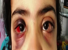
Citation: Salahiddine Saghir, Zainab Rifai, Rachid Abilkassem, et al. Ocular Manifestations of Acute Myeloid Leukemia. Clin Image Case Rep J. 2021; 3(1): 128.

Ocular Manifestations of Acute Myeloid Leukemia
Abstract

A 5-year-old Infant, followed for trisomy 21 and treated for Two years for acute myeloid leukemia, has been admitted with visual disturbances and ocular swelling. The physical examination showed oculomotor paralysis, conjunctival hemorrhage, periorbital swelling and bruising (Figure 1), gingival hypertrophy was also noticed (Figure 2). A fundus examination showed Viscosity-related retinopathy with white center hemorrhage. The orbital scan performed revealed a thickening of the soft parts of the periorbital region.

The blood screening and the blood smear showed an invasion of the peripheral blood by the blasts, allowing the diagnosis of recurrent leukemia. the patient was referred to the pediatric oncology center for therapeutic treatment.
Ocular involvement is the third extra-medullary location after Central Nervous System and testicular involvement. Several lesions remain asymptomatic and will be diagnosed after an ophthalmologic examination of routine [1].
All the parts can be affected, the first sign of anterior segment involvement during leukemia is iris heterochromia or nodules, uveitis with hypopyon, isolated ocular hypertonia, or spontaneous hyphemia. The retina is very commonly affected. It is estimated that over 70% of patients have retinal abnormalities during ocular relapse. The early retinal manifestations are venous dilations and tortuosities [2].

Early detection of subclinical ophthalmologic lesions is crucial, the ophthalmological examination must be systematic, completed by paraclinical examinations. It is still much more important in patients in complete remission, the aim being to detect relapses as soon as possible, as early treatment can improve the life expectancy of the patient.