Clinical Images and Case Reports Journal | PubMed
  • contact@literaturepublishers.org
  • Whitley Bay, NE26 2HU, England, UK
  • Submit Manuscript
Article Details

Clinical Image

Volume 3, Issue 1 (January Issue)

Primary Localized Giant Adrenal Cortical Carcinoma in a Young Adult Woman

Rosita Sortino1*, Michael Schmid2, Luca Benigno1 and Walter Kolb1

1Department of Surgery, Kantonsspital St. Gallen, St. Gallen, Switzerland
2Department of Pathology, Kantonsspital St. Gallen, St. Gallen, Switzerland

*Corresponding author: Dr. Med. Rosita Sortino, Department of Surgery, Cantonal Hospital St. Gallen Rorschacherstrasse, St. Gallen, Switzerland, Tel: +41714949314; E-mail: rosita.sortino@kssg.ch

Received: November 26, 2020; Accepted: December 14, 2020; Published: January 03, 2021

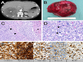
Citation: Rosita Sortino, Michael Schmid, Luca Benigno, et al. Primary Localized Giant Adrenal Cortical Carcinoma in a Young Adult Woman. Clin Image Case Rep J. 2021; 3(2): 134.

Primary Localized Giant Adrenal Cortical Carcinoma in a Young Adult Woman
Abstract

A previously healthy 45-year-old woman was referred to our Department of Surgery with a three-month history of left flank pain, weight loss and intermittent fever associated with a large mass of the right adrenal gland discovered sonographically by the patient’s general practitioner. Physical examination showed no abnormalities. An unremarkable biochemical profile (aldosterone, cortisol, metanephrin, renin, sexual hormone) accompanied by constant normal clinical signs ruled out pheochromocytoma.

Abdominal Computed Tomography (CT) scans (Figure 1A) demonstrated a solid mass in the right adrenal gland with an average diameter of 12 cm. No distant metastases were found in the staging workup (stage II ACC). Due to the patient’s young age and the radiologic absence of metastatic disease our multidisciplinary team decided on surgical resection and the patient underwent complete open right adrenalectomy (R0). On gross examination, the enlarged adrenal gland (16 x 11 x 10 cm, 980 g; Figure 1B) showed an encapsulated, multilobulated, partially necrotic tumor measuring 12 cm in diameter. Microscopic examination revealed a nodular proliferation of highly pleomorphic cells separated by fibrous bands and multiple areas of necrosis with increased mitotic activity as well as capsular and lymphovascular invasion.

Immunohistochemical analysis revealed strong and diffuse positivity for Inhibin-? and Synaptophysin, focal positivity for Vimentin and Melan A, and an increased proliferation fraction (Ki-67) of 40%. Based on morphology and immunohistochemistry, the diagnosis of adrenal cortical carcinoma, high-grade, was established. The patient was discharged four days after surgery. Considering the high Ki-67 expression, systemic adrenalitic chemotherapy with mitotane with curative intent for three years was recommended. Follow-up CT 12 months after surgery showed no signs of local recurrence.