
Clinical Images and Case Reports Journal | PubMed
  • contact@literaturepublishers.org
  • Whitley Bay, NE26 2HU, England, UK
  • Submit Manuscript
Article Details

Case Report

Volume 2, Issue 3 (August Issue)

Pulmonary Typical Carcinoid on Right Middle Lobe Hypoplasia: A Case Report and Review of the Literature

Nozomu Motono1*, Akihiko Shioya2, Sohsuke Yamada2 and Hidetaka Uramoto1

1Department of Thoracic Surgery, Kanazawa Medical University, 1-1 Daigaku, Uchinada, Ishikawa, 920-0293, Japan

2Department of Clinical Pathology, Kanazawa Medical University, 1-1 Daigaku, Uchinada, Ishikawa, 920-0293, Japan

*Corresponding author: Nozomu Motono, Department of Thoracic Surgery, Kanazawa Medical University, 1-1 Daigaku, Uchinada, Ishikawa, 920-0293, Japan, E-mail: motono@kanazawa-med.ac.jp

Received: July 22, 2020; Accepted: August 03, 2020; Published: August 25, 2020

Citation: Motono N, Shioya A, Yamada S, et al. Pulmonary Typical Carcinoid on Right Middle Lobe Hypoplasia: A Case Report and Review of the Literature. Clin Image Case Rep J. 2020; 2(3): 120.
Pulmonary Typical Carcinoid on Right Middle Lobe Hypoplasia: A Case Report and Review of the Literature
Abstract

Introduction: Pulmonary hypoplasia is common in the perinatal period and a significant cause of death in newborn infants. The majority of patients present with severe respiratory distress or repeated pulmonary infections. However, there have been some cases of pulmonary hypoplasia being diagnosed in adulthood. Furthermore, there have been some reports of lung cancer and Diffuse Idiopathic Pulmonary Neuroendocrine Cell Hyperplasia (DIPNECH) developing in pulmonary hypoplasia patients.

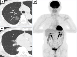
Case Presentation: A 70-year-old man was aware of dry cough. Chest computed tomography showed a ground-glass nodule in the upper lobe of the right lung and middle lobar incomplete development of the right lung. Lung cancer of the right upper lobe and pulmonary hypoplasia of the right middle lobe was suspected, and right upper and middle lobectomy and lymph node dissection were performed. A pathological examination confirmed lepidic adenocarcinoma of the right upper lobe and a well-differentiated neuroendocrine tumor with an organoid or trabecular pattern at the middle lobe of the right lung. Based on the results of an immunohistochemical study, the tumor of the middle lobe was diagnosed as typical carcinoid.

Conclusion: We experienced a rare case of pulmonary typical carcinoid in a patient with middle lobe hypoplasia of the right lung coexisting with lung adenocarcinoma on the upper lobe of the right lung.

Keywords: Pulmonary hypoplasia; Carcinoid; Lung cancer; Diffuse idiopathic pulmonary neuroendocrine cell hyperplasia

hacklink satın al coinbar coinbar giriş grandpashabet grandpashabet giriş grandpashabet güncel giriş grandpashabet grandpashabet giriş grandpashabet giriş güncel alobet alobet giriş betcio betcio giriş betcio betcio giriş holiganbet giriş holiganbet jojobet jojobet giriş meritking meritking giriş meritking meritking giriş meritking giriş meritking jojobet güncel giriş best online casinos in new zealand holiganbet holiganbet giriş holiganbet güncel giriş holiganbet holiganbet giriş holiganbet güncel giriş betcio betcio giriş alobet alobet giriş atlasbet atlasbet giriş pashagaming pashagaming giriş pashagaming pashagaming giriş madridbet madridbet giriş kingroyal kingroyal giriş holiganbet holiganbet giriş grandpashabet grandpashabet giriş grandpashabet güncel giriş holiganbet holiganbet giriş holiganbet güncel giriş atlasbet atlasbet giriş atlasbet güncel giriş holiganbet betbey esbet holiganbet jojobet holiganbet jojobet giriş holiganbet 1win cashwin betpuan sonbahis grandpashabet casinoroyal grandpashabet jasminbet jasminbet giriş meritking meritking giriş meritking güncel giriş meritking meritking giriş meritking güncel giriş pashagaming pashagaming giriş pashagaming güncel giriş betcio betcio giriş betcio güncel giriş