Clinical Images and Case Reports Journal | PubMed
  • contact@literaturepublishers.org
  • Whitley Bay, NE26 2HU, England, UK
  • Submit Manuscript
Article Details

Clinical Image

Volume 3, Issue 3 (March Issue)

Rare Examples of the Space of Okada Spread

Yakov Perper*

Department of Anesthesiology, Mount Sinai Hospital of Queens, Queens, NY, USA

*Corresponding author: Yakov Perper, Department of Anesthesiology, Mount Sinai Hospital of Queens, Queens, NY, USA, E-mail: yperper@universalpainmanagement.nyc

Received: February 05, 2021; Accepted: February 13, 2021; Published: March 24, 2021

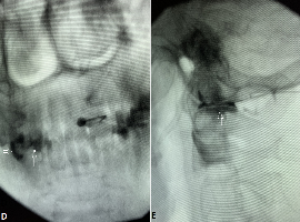
Citation: Yakov Perper, et al. Rare Examples of the Space of Okada Spread. Clin Image Case Rep J. 2021; 3(3): 146.

Rare Examples of the Space of Okada Spread
Abstract

I combined two cases of the contralateral facet contrast distribution as they are quite rare. In the first case (Figure 1A, B and C), it was a cervical epidural injection, and in the second (Figure D and E), it was an Atlanto-Axial (AA) joint injection. The current explanation of these phenomena is the retrodural Space of Okada (SOO) spread [1,2]. First described as the space providing communication between the contarlateral facet joints [1], SOO distribution with AA joint injection (Figure D and E) is very unusual. It is the only case in my practice for many years. However, I frequently see the SOO opacification with my cervical epidural injections [3]. Most of the time, it is ipsilateral, randomly bilateral, and as an exception, only contralateral (Figure A, B and C). I frequently encounter SOO with cervical ESIs due to the technique I employ. Instead of the radiolucent air or saline traditionally used with loss of the resistance technique, I inject contrast and carefully explore the resulting images [4]. Treated by some as a complication, SOO opacification is actually a normal flow pattern, and further needle advancement is required for epidural access (Figure C) [3]. It is unclear if the pathway for the contralateral contrast distribution is the same with the epidural (Figure A, B and C) and intraarticular (Figure D and E) injections. Future research is warranted for a better understanding of the nature of the space of Okada.