Clinical Images and Case Reports Journal | PubMed
  • contact@literaturepublishers.org
  • Whitley Bay, NE26 2HU, England, UK
  • Submit Manuscript
Article Details

Clinical Video

Volume 8, Issue 2

Space-Occupying Cerebellar Infarction: Intraoperative Visualization of Raised Intracranial Pressure and Cerebellar Tissue Prolapse

Daniel Schöni1* and Alex Alfieri1,2

1Department of Neurosurgery, Cantonal Hospital of Winterthur, Winterthur, Switzerland
2Neurocenter of Southern Switzerland, Faculty of Biomedical Sciences, University of Southern Switzerland, Lugano, Switzerland

*Corresponding author: Daniel Schöni, Department of Neurosurgery, Cantonal Hospital of Winterthur, Winterthur, Switzerland. E-mail: daniel.schoeni@ksw.ch

Received: April 06, 2026; Accepted: April 20, 2026; Published: May 05, 2026

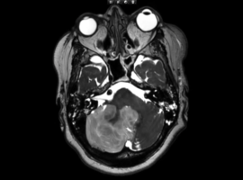
Citation: Schöni D, Alfieri A. Space-Occupying Cerebellar Infarction (SOCI): Intraoperative Visualization of Raised Intracranial Pressure and Cerebellar Tissue Prolapse. Clin Image Case Rep J. 2026; 8(2): 588.

The following video is related to this article (Video 1).

Space-Occupying Cerebellar Infarction: Intraoperative Visualization of Raised Intracranial Pressure and Cerebellar Tissue Prolapse
Abstract

A 60-year-old woman underwent emergency replacement of the ascending aorta and aortic hemiarch with a supracoronary tube graft for acute type A intramural hematoma. The postoperative course was critical and complicated by neurological deterioration. Initial brain CT demonstrated a right PICA territory infarction. Progressive neurological worsening culminating in a comatose state prompted additional MRI, which revealed progression of the space-occupying cerebellar infarction (Figure 1).
To alleviate increased pressure within the posterior fossa, a navigated mini-osteoplastic suboccipital craniotomy with partial resection of infarcted cerebellar tissue was performed. Upon dural opening, immediate prolapse of infarcted cerebellar tissue confirmed increased intracranial pressure (Video 1).
Following neurosurgical intervention, the patient showed rapid neurological improvement. Within five days, she regained the ability to communicate and demonstrated full movement of all extremities. Follow-up CT imaging confirmed adequate posterior fossa decompression, a distinct resection cavity, and relief of brainstem compression (Figure 2).

Clinical Video
Video showing the intraoperative view of a small (2 × 2 cm) right-sided posterior fossa craniotomy with the dura initially closed. Upon opening the dura, immediate cerebellar tissue prolapse is observed due to increased intracranial pressure.