Clinical Images and Case Reports Journal | PubMed
  • contact@literaturepublishers.org
  • Whitley Bay, NE26 2HU, England, UK
  • Submit Manuscript
Article Details

Clinical Image

Volume 4, Issue 7 (July Issue)

Stewart-Traves Syndrome

Wen-Hsien Hsu1* and Wei-Yu Chen2

1Division of Lymph-vascular Surgery, Department of Surgery, Wan Fang Hospital, Taipei Medical University, Taiwan

2Department of Pathology, Wan-Fang Hospital, Taipei Medical University, Taiwan

*Corresponding author: Wen-Hsien Hsu, Division of Lymph-vascular Surgery, Department of Surgery, Wan Fang Hospital, Taipei Medical University, Taiwan. E-mail: angiohsu@gmail.com

Received: June 25, 2022; Accepted: July 03, 2022; Published: July 20, 2022

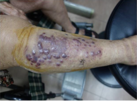
Citation: Hsu WH, Chen WY. Stewart-Traves Syndrome. Clin Image Case Rep J. 2022; 4(7): 246.

Stewart-Traves Syndrome
Abstract

The Stewart-Traves syndrome is a rare and deadly entity, which is defined as angiosarcoma arising in the setting of chronic lymphedema. Originally STS is referring to the woman who develop lymphedema in the upper extremity secondary to axillary lymph node dissection for breast cancer. It is extremely rare in the lower extremity because of secondary lymphedema.

An eighty-seven-year-old lady, a native of Taiwan, presented herself to the lymph-vascular clinic with complaints of chronic swollen right leg with newly developed blisters in the mid-portion of right leg. The patient needs ambulatory assistance due to pain and tight sensation in the affected lower limb. Her past history revealed radical hysterectomy in the remote past for cervical cancer. No radiotherapy or chemotherapy reported in the postoperative course. For 5 years she has been experiencing swollen right lower limb with recurrent bouts of cellulitis. The 3-dimention image study showed bilateral iliac vein compression, for which She underwent angioplasty with iliac vein stenting. The swollen right leg then temporarily improved, but newly developed multiple purple bullae was formed.

Skin biopsy was then performed. The pathology report showed (Figure 1) epithelioid angiosarcoma with epithelioid tumor cells marked nuclear pleomorphism and prominent nucleoli (Figure 2) angiosarcoma with anastomosing vascular structures dissecting collagen fibers (Figure 3). Positive CD31 immunostain in the tumor cells. Due to patient’s poor condition, further staging workup and possible chemotherapy were declined by family.