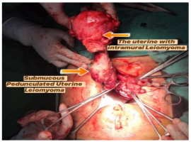
A 40-year-old woman from Indonesia a history of heavy menstrual bleeding sometimes with clots and irregular per vagina bleeding since one month ago. She reported low abdominal pain, abdominal dilated and pelvic pain. Speculum examination showed a round-shaped mass amount duck's egg out of external uterine ostium like had pedunculated from the uterine cavity and showed blood on the cervical canal. On vaginal toucher examination showed Fluxus, a round-shaped mass amount duck's egg was palpable, slippery surface, smooth, not weak, not slinger pain. The size of the corpus uterine as big as a fist. Submucous uterine leiomyoma is common benign tumors in the wall uterus, which are present in approximately 15-20?ses and it rarely prolapse into the vagina through the cervical canal, especially when it had pedunculated.
Patients with uterine leiomyoma, generally present with symptoms of irregular and heavy menstrual bleeding, pelvic pain, pressure effects, and anemia. In this case, the patient presented a history of irregular and heavy menstrual bleeding since one month ago, lethargy, low abdominal pain, abdominal dilated and pelvic pain. In our case, we diagnosed the patient with anamnesis, physical examination, speculum examination, vaginal toucher and hematological examination. In this case, the uterus had two cases was uterine leiomyomas which are submucous pedunculated uterine leiomyoma and intramural leiomyoma. So, that is the reason why we did a total abdominal hysterectomy for the patient.
")
")