
Clinical Images and Case Reports Journal | PubMed
  • contact@literaturepublishers.org
  • Whitley Bay, NE26 2HU, England, UK
  • Submit Manuscript
Article Details

Clinical Image

Volume 3, Issue 8 (August Issue)

Unusual Presentation of Malignant Breast Tumor

Fariba Yazdanpanah1*, Gurdeep Chhabra2, Tekabe Nigussie Birhane1, Sarwat Waqar Siddiqui3 and Ellen Manlucu3

1Internal Medicine Department, University of Maryland Capital Region Medical Center, USA
2Hematology/Oncology Department, University of Maryland Capital Region Medical Center, USA
3Pathology Department, University of Maryland Capital Region Medical Center, USA

*Corresponding author: Fariba Yazdanpanah, Internal Medicine Department, University of Maryland Capital Region Medical Center, USA. E-mail: Fariba.Yazdanpanah@umm.edu

Received: August 30, 2021; Accepted: September 06, 2021; Published: September 16, 2021

Citation: Yazdanpanah F, Chhabra G, Birhane TN, et al. Unusual Presentation of Malignant Breast Tumor. Clin Image Case Rep J. 2021; 3(8): 180.
Unusual Presentation of Malignant Breast Tumor
Abstract

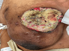
A 78-year-old, African American, legally blind female presented to the emergency department following recurrent mechanical falls. The patient’s past medical history was significant for atrial fibrillation, diabetes mellitus, hypertension, and hyperlipidemia and her family history was remarkable for ovarian cancer in her sister and prostate cancer in her brother. On physical exam, a large, necrotic, fungating, right breast mass with purulent drainage was found (Figure 1). The onset, per patient, was 4-5 months prior to presentation and the patient never had it evaluated. Chest computed tomography (CT) scan with contrast revealed a large ulcerating right breast tumor (14 x 6 x 11 cm) with infiltration of the right chest wall musculature (Figure 2), large right axillary lymph node metastases, and multiple pulmonary metastatic lesions (compatible with a stage T4b N3 M1 lesion). The patient underwent a core-needle biopsy, and the report showed invasive ductal carcinoma grade 2 (Figure 3). The immunohistochemical stain for E-cadherin was positive confirming the ductal phenotype (Figure 4). As per the current College of American Pathologists (CAP) and American Society of Clinical Oncology (ASCO) criteria, hormone receptor testing was done and resulted with Estrogen Receptor (ER): Negative, Progesterone Receptor (PgR): Positive (Percentage of cells with nuclear positivity: less than 5%), and Human Epidermal growth factor Receptor 2 (HER2): Negative. The patient was considered for systemic chemotherapy and breast-conserving therapy (BCT) via radiation therapy to palliate symptoms, prolong survival, and maintain quality of life.

hacklink satın al casibom jojobet jojobet giriş deneme bonusu deneme bonusu veren siteler betsat betsat giriş betsat güncel giriş deneme bonusu deneme bonusu veren siteler perabet perabet giriş milanbahis milanbahis giriş holiganbet holiganbet giriş holiganbet holiganbet giriş jojobet jojobet giriş casibom giriş holiganbet holiganbet giriş holiganbet güncel giriş perabet perabet giriş perabet güncel giriş perabet perabet giriş perabet güncel giriş casibom casibom giriş casibom güncel giriş meritking meritking giriş meritking güncel giriş holiganbet holiganbet giriş holiganbet güncel giriş jojobet jojobet giriş jojobet güncel giriş kingroyal kingroyal giriş kingroyal güncel giriş bahsegel bahsegel bahsegel marsbahis jojobet marsbahis marsbahis bahsegel bahsegel egebet Egebet güncel giriş jojobet jojobet giriş nakitbahis nakitbahis giriş nakitbahis güncel giriş nakitbahis nakitbahis giriş Nakitbahis nakitbahis giriş nakitbahis güncel giriş jojobet jojobet giriş perabet perabet giriş bahsegel casibom giftcardmall/mygift betcio betcio giriş betcio güncel giriş padişahbet padişahbet giriş casibom casibom casibom jojobet jojobet jojobet perabet perabet giriş