Clinical Images and Case Reports Journal | PubMed
  • contact@literaturepublishers.org
  • Whitley Bay, NE26 2HU, England, UK
  • Submit Manuscript
Article Details

Clinical Image

Volume 7, Issue 5

Diagnostic Dilemma: Evolving Nodules in a Giant Congenital Melanocytic Nevus

Sophia Manduca1 and Genevieve Kaunitz2*

1New York University Grossman School of Medicine, New York, NY, USA
2Department of Dermatology, University of California San Diego School of Medicine, San Diego, CA, USA

*Corresponding author: Genevieve Kaunitz, MD, Department of Dermatology, University of California San Diego School of Medicine, San Diego, CA, USA.
E-mail: gkaunitz@health.ucsd.edu

Received: July 23, 2025; Accepted: August 06, 2025; Published: August 15, 2025

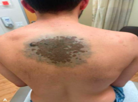
Citation: Manduca S, Kaunitz G. Diagnostic Dilemma: Evolving Nodules in a Giant Congenital Melanocytic Nevus. Clin Image Case Rep J. 2025; 7(5): 568.

Diagnostic Dilemma: Evolving Nodules in a Giant Congenital Melanocytic Nevus
Abstract

A 29-year-old man presented for a skin examination and noted development of new, tender nodules within a congenital birthmark on his upper back. Physical examination demonstrated a 14 by 12 cm homogenous blue patch, with numerous central brown macules coalescing into patches with scattered blue-black thin papules within. The largest was a 1 cm blue-black plaque on the left lateral edge of the lesion. A shave biopsy of the tender lesion showed a giant congenital melanocytic proliferation with features of Masson’s neuronevus. Given ongoing clinical suspicion for malignancy, two subsequent punch biopsies of additional nodules were performed, which showed similar findings with features overlapping those of an epithelioid blue nevus.
Giant congenital melanocytic nevi are a recognized risk factor for melanoma, with estimated lifetime risk ranging from 5-40% [1]. In this patient, the emergence of new nodules and evolving pigmentation raised clinical suspicion for malignant transformation. However, histopathologic evaluation confirmed a benign melanocytic process. The lesion remained stable and asymptomatic on follow-up.
This image underscores the diagnostic challenge posed by evolving lesions in giant congenital melanocytic nevi and highlights the critical role of biopsy in distinguishing benign proliferative nodules from melanoma in this high-risk population.